Gallery
Annotated examples, training images, and reference materials organized by dataset and category.
BANC
Brain and Nerve CordFull brain and ventral nerve cord dataset providing complete CNS connectivity.
 Structure
Soma Arrangement (Color Overlay)
Dense cluster of cell bodies with color segmentation overlay. Each unique color represents an individually segmented soma.
 Structure
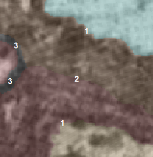
Single Soma (Color Overlay)
Individual neuronal soma highlighted with color segmentation overlay, showing the cell body boundary and surrounding neighbors.
 Structure
Dense Soma Region
Region densely packed with neuronal cell bodies. Nuclei and surrounding neuropil visible throughout.
 Structure
Sparse Soma Region
Region with fewer, well-separated cell bodies surrounded by extensive neuropil.
 Structure
Dark Soma (Possible Glia)
Darkly stained cell body with electron-dense cytoplasm, possibly glial in origin. Note the distinct contrast difference from surrounding neurons.
 Structure
Large Soma with Organelles
Exceptionally large neuronal soma rich in organelles including prominent nucleus, endoplasmic reticulum, and mitochondria.
 Structure
Large Soma with Organelles (View 2)
Second perspective of a large soma showing dense organelle packing. Note the extensive rough endoplasmic reticulum and clustered mitochondria.
 Structure
Large Soma with Organelles (View 3)
Third view highlighting the scale of a large neuronal soma relative to surrounding neurites and neuropil.
 Structure
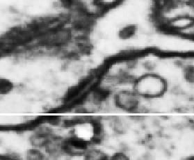
Possibly Myelinated Soma
Soma with an unusually thick membrane border, potentially myelinated. Note: horizontal line artifact from monitor capture is also visible.
 Structure
Thick Membrane Border
Soma enclosed by an unusually thick, electron-dense membrane layer. Possible myelination of a cell body — an uncommon but documented feature. Is this a thing?
 Structure
Golgi Apparatus and Organelles
Golgi apparatus visible alongside mitochondria, cisternae, microvesicles, and vacuoles near a soma. Cell membrane border clearly delineated.
 Structure
Golgi Apparatus (Detail)
Close-up view of Golgi apparatus showing stacked cisternae and associated vesicles within the cytoplasm.
 Structure
Membrane Whorls and Golgi Apparatus
Large, heavily stained membrane whorls alongside Golgi apparatus and various organelles within or near a cell body.
 Structure
Dense Organelle Cluster Near Soma
Heavily stained organelles including mitochondria clustered near a soma. Note the high electron density contrasting with surrounding cytoplasm.
 Structure
Membrane Whorl
Concentric membrane whorl forming a spiral pattern. These lamellar structures are occasionally encountered and may represent myelin figures or autophagic bodies.
 Structure
Membrane Spirals (Close-up)
High-magnification view of concentric membrane spirals. The tightly wound lamellae create a distinctive fingerprint-like pattern.
 Structure
Orphan Mitochondria Cluster
Isolated cluster of mitochondria and heavily stained myelin-like material. Appears to be an orphan object disconnected from a parent structure.
 Structure
Orphan Mitochondria Cluster (With Context)
Same orphan cluster shown with surrounding neuropil, dataset coordinates, and layer information for reference.
 Structure
Heavily Stained Dark Organelles
Multiple organelles with abnormally heavy staining producing high-contrast, electron-dense profiles. Green horizontal line is a monitor capture artifact. Not sure where that comes from, hmm.
 Structure
Unidentified Dense Body
Dense, heavily stained structure of uncertain identity — possibly an organelle or a localized section artifact. Differential diagnosis is challenging at this magnification.
 Structure
Lipid Droplets
Cluster of lipid droplets (fat globules) appearing as electron-dense spherical inclusions. Commonly found near cell bodies.
 Structure
Staining Anomaly with Micro Tear
Heavily stained structure with irregular contrast and a small tear visible within a strange organelle. Combines staining artifact with minor physical damage.
 Structure
Parallel Axon Bundle
Bundle of neurites sectioned in the parallel plane, probably axonal. Note the uniform elongated profiles and consistent diameter.
 Defect
Section Damage
Physical damage to the tissue section visible as a dark linear disruption. Green arrows indicate affected regions. Do not annotate across damaged areas.
 Defect
Missing Section
Gap in tissue where a slice is absent, creating a void (black region). Green arrows point to the boundary. Data is unrecoverable in these areas.
 Defect
Membrane Blowout
Localized membrane blowout where tissue has ruptured outward, creating a washed-out void. Green outline marks the affected boundary.
 Defect
Section Pinch
Tissue compression artifact where the section was pinched during preparation, creating artificial convergence of structures.
 Defect
Section Stretch
Stretching artifact showing tissue pulled along one axis. Green arrows indicate the direction of distortion. Structures appear elongated and thinned.
 Defect
Section Tear
Linear tear through the tissue section creating a clean void. Green arrows mark the tear boundaries. Do not trace structures across the gap.
 Defect
Resin Tear
Tear occurring within the embedding resin rather than the tissue itself. Shows clean break edges characteristic of resin fracture.
 Defect
Section Folds (Overview)
Zoomed-out view of the central brain region showing prominent section folds crossing the tissue. Dark diagonal lines are folded-over tissue creating doubled layers.
 Defect
Folds with Rippling
Zoomed-out view showing section folds accompanied by wavy, faded rippling patterns. Tissue appears distorted across a large area.
 Defect
Severe Section Folds
Extreme folding artifact with multiple overlapping fold layers creating near-total data loss in affected regions.
 Defect
Staining Artifact
Uneven heavy metal staining producing localized dark deposits that obscure underlying ultrastructure.
 Defect
Splotch Artifact
Irregular dark splotching at section boundaries, likely from staining contamination or resin pooling during preparation.
 Defect
Combined Defects: Staining, Fold, Lipid Droplets
Multiple artifacts in one region: uneven staining, tissue fold (dark diagonal band), and scattered lipid droplets (fat globules).
 Defect
Lipid Droplets (Section Artifact)
Scattered lipid droplets (fat globules) appearing as dark spherical inclusions. Green arrows highlight individual droplets. Can be confused with biological structures.
 Defect
Multiple Section Errors (Central Chiasm)
Central chiasm region exhibiting multiple simultaneous defects: large tissue distortions, folds, and torn areas. Severely compromised data integrity.
 Defect
Section Cracks (aka Lightning™)
Zoomed-out view showing branching cracks propagating across the section. Nicknamed “lightning” for their characteristic branching pattern.
 Defect
Multiple Section Errors (Overview)
Panoramic view showing a convergence of defect types: folds, cracks, pinch artifacts, and a missing section — all in one region.
FAFB 2019
FlyWireFull Adult Fly Brain dataset. Primary source for Drosophila connectomics research.
 Synapse
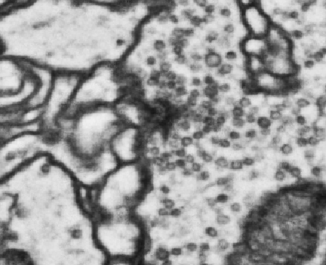
T-bar Synapse
Classic T-bar morphology with platform structure, dense body visible in cross-section.
 Synapse
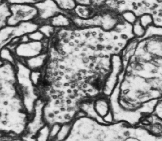
Elongated T-bar
Extended platform variant. Note the longer horizontal extent compared to standard T-bars.
 Synapse
Multiple T-bars (Smudge)
Clustered synapses appearing as smudge-like shapes. Common in regions of high synaptic density.
Reference Materials
TrainingProtocol reference images and training materials from expert annotators.
 Synapse
GT Protocol Reference 6
Ambiguous cases and decision criteria for edge cases.
Image Bounty List
WantedWe're looking for good examples of these structures and phenomena. Have an image you want to share? Submit it on GitHub.